
The simultaneous presence of blue and yellow in the iris involves distinct pigmentary mechanisms. Distinguishing a simple gradient of melanin from a pathological signal requires precisely locating the yellow hue: iris, sclera, or conjunctiva tell different clinical stories.
Heterogeneous pigmentation of the iris: melanin and lipochrome
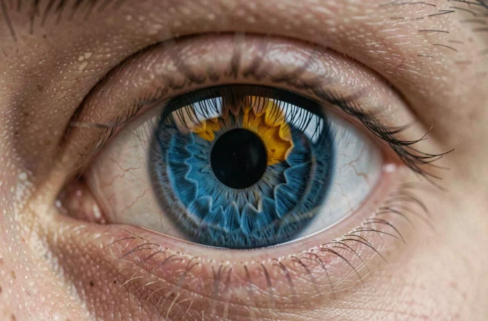
A blue iris indicates a low concentration of melanin in the iris stroma. Incident light undergoes Rayleigh scattering on collagen fibers, reflecting a short wavelength perceived as blue. No blue pigment exists in the human iris.
The yellow or golden component sometimes observed around the pupil comes from a distinct pigment, lipofuscin (often referred to as lipochrome in the ocular context). This deposit concentrates in the peripupillary area and creates a hazel to golden ring against a blue background.
We frequently observe this combination in individuals of European descent carrying variants of the OCA2 gene associated with reduced melanin production. The coexistence of blue and yellow in the iris has no pathological value in itself. It reflects an intermediate pigment dosage, sometimes referred to as “hazel” or cool dominant hazel eyes. When discussing blue and yellow eye color, it is most often this benign pigment mosaic that is involved.
Yellow sclera and bilirubin: the hepatic signal not to be confused
The real medical issue concerns the sclera, not the iris. Yellowing of the white of the eye indicates an accumulation of bilirubin in the blood, a condition known as jaundice. Bilirubin is a breakdown product of hemoglobin, normally conjugated by the liver and then excreted in bile.
When the liver malfunctions, when the bile ducts are obstructed, or when excessive hemolysis releases too much bilirubin, the pigment accumulates in elastin-rich tissues, primarily the sclera. This is why scleral jaundice is often the first visible sign, even before skin yellowing.
Common causes of scleral jaundice
- Liver damage: viral hepatitis, advanced steatosis, cirrhosis, drug-induced hepatitis. The liver can no longer effectively conjugate bilirubin.
- Biliary obstruction: choledocholithiasis, tumor of the head of the pancreas, or cholangitis. Bile no longer flows to the duodenum and backs up into the blood.
- Pathological hemolysis: autoimmune hemolytic anemias, sickle cell disease, G6PD deficiency. The accelerated destruction of red blood cells saturates the liver’s conjugation capacity.
A diffuse yellowing of the sclera is always a reason for prompt consultation. A common mistake is to attribute it to fatigue or poor lighting.
Localized yellow eye or diffuse jaundice: distinguishing criteria
Not every yellowish hue on the eye is related to jaundice. We recommend checking three parameters before becoming alarmed.
The first is localization. A yellowish deposit limited to the bulbar conjunctiva, often nasal, suggests a pinguecula. This benign degenerative lesion of conjunctival tissue is linked to chronic UV exposure and dryness. It does not indicate any liver dysfunction.
The second is bilaterality. Scleral jaundice is always bilateral and symmetrical. Unilateral or focal coloring points towards a surface pathology (pinguecula, early pterygium, dermoid cyst).
The third is the clinical context. Jaundice is almost always accompanied by associated signs:
- Dark urine (tea or brown beer color), indicating renal excretion of conjugated bilirubin
- Pale stools (light-colored, clay-like), signaling the absence of stercobilin in the digestive tract
- Pain in the right upper quadrant, fever, generalized itching, or confusion in severe forms
- Persistent nausea or vomiting without an obvious digestive cause
In the absence of these signs, a localized yellow reflection on the conjunctiva is rarely concerning.
Blue eyes and specific ocular risks
Light-colored irises allow more light to reach the retina. The low density of iris melanin reduces UV filtering and blue wavelengths. This characteristic has practical consequences.
The HUG Foundation classifies light eyes among the factors promoting the onset of age-related macular degeneration (AMD), alongside heredity, smoking, and overweight. Therefore, sun protection through UV-filtering sunglasses is more crucial for individuals with blue irises than for those with densely pigmented irises.
Light sensitivity (functional photophobia) is also more pronounced in individuals with blue eyes. It is not a pathology but justifies wearing tinted or photochromic lenses in bright conditions.
Recommended protection and follow-up
Wearing UV 400 filter sunglasses remains the simplest measure. For contact lens wearers, some contact lenses now incorporate a UV filter of class 1 or 2, although they do not replace wraparound sunglasses.
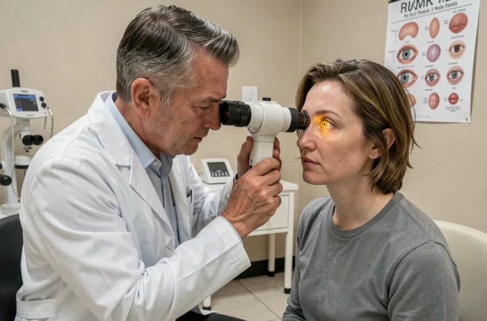
A regular fundus examination after the age of fifty allows for the detection of early signs of AMD (drusen, pigmentary changes) at a stage where treatments remain effective.
The blue-yellow combination in the iris remains a pigmentary trait without diagnostic value. The only yellow that deserves medical attention is that which diffusely and bilaterally colors the sclera, especially when accompanied by dark urine or abdominal pain. Confusing the two means either worrying for nothing or ignoring a hepatic signal that requires a blood test in the following days.